PLEURAL FLUID TAPPING
Pleural Fluid Tapping / Thoracocentesis / Pleural Aspiration
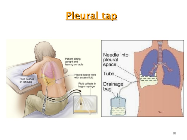
Image Source: https://www.slideshare.net/rongon28us/thorax-pleura
This is a procedure to remove fluid from the space between the lining of the outside of the lungs (pleura) and the wall of the chest. Normally, very little fluid is present in this space. An accumulation of excess fluid between the layers of the pleura is called a pleural effusion.
This test is generally done by the patients’ bedside in the intensive care unit. A chest radiograph would hint towards the formation of an effusion. Using Ultrasound guidance the site and size of the effusion is assessed. The area is then marked and the site is prepared for the procedure using cleaning material to minimise the risk of infection.
A local anaesthetic, or numbing agent, is then given underneath the skin and around the area of the procedure. A needle is then placed through the skin of the chest wall into the space around the lungs called the pleural space. Fluid is withdrawn and collected and may be sent to a laboratory for analysis (pleural fluid analysis).
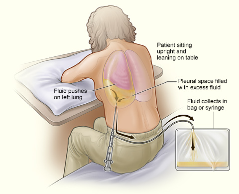
Image Source: https://en.wikipedia.org/wiki/Thoracentesis
When done under imaging guidance, the test is relatively safe, however there is a small risk of possible complications. This includes:
Pneumothorax (collapse of the lung)
Fluid re-accumulation
Pulmonary oedema
Bleeding
Infection
Respiratory distress


Ask a Question?