RADIOLOGY
IMAGING IN ICU SET-UP
Radiological imaging is an integral part of management in ICU set up. The commonly used modalities are X Ray, USG, CT scan and MRI.
X- Ray or Rhontgenogram
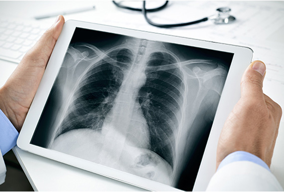
Image Source: https://www.medicalnewstoday.com/articles/153201.php
X- Ray or Rhontgenogram is a basic imaging modality which gives a 2D representation of the body part to be evaluated. In an ICU set-up, X Rays are taken by portable X Ray machine that can be placed next to the patient, hence avoiding the need to shift the patient out of the ICU. The most common X Ray taken is a Chest X Ray. It is advised by the doctor for ICU patients to evaluate disease progression or remission. It may be asked on a daily basis, in case the patient condition is critical or as may be clinically indicated. It is mandated in cases of insertion of various tubes and catheters, to look for their satisfactory position.
USG

Image Source: http://welkinmedicare.com/services/diagnostic/imaging/usg
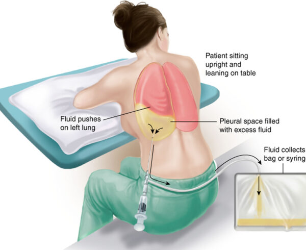
USG is also an effective bedside imaging modality that makes use of ultrasonic sound waves for imaging. There are no ionizing radiations in USG. It is advised by the doctor for disease under evaluation, most commonly for abdomen. It is also helps in placement of various lines and catheters. In patients with fluid in abdomen or chest, USG guided tapping(drainage) of fluid may be done. Sometimes a Doppler of lower limb or upper limb may be indicated in case of swelling, to rule out deep venous thrombosis.
CT Scan
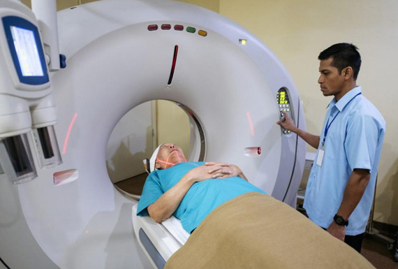
Image Source: https://www.medicalnewstoday.com/articles/153201.php
CT scan is a very useful imaging modality and gives invaluable information of the disease under evaluation. The scan takes short time for imaging and detailed information can be obtained. It is commonly used in to look for a focus of suspected infection, in case of bleeding, brain hemorrhage, raised intracranial pressure (due to many causes), brain infarct, bowel obstruction, perforation, etc; all of which require immediate attention and appropriate treatment.
MRI
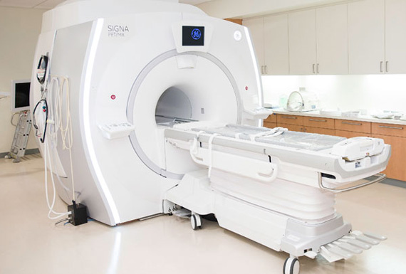
Image Source: https://stanfordhealthcare.org/medical-tests/p/pet-mri-scan.html
MRI is a more advanced modality which is most commonly used in an ICU set-up for brain imaging. Other body parts may also be imaged depending upon the clinical indication. MR imaging takes a long time and is always supervised by a qualified anaesthetist for all ICU patients. It does not make use of any ionising radiation. It makes use of magnetic field to generate high quality images and thereby aiding in diagnosis and management.
FAQ's
Is X Ray radiation harmful?
The radiation dose of single chest X Ray is on an average 0.1 to 0.2mSv and as small as 0.01 for extremities, which is a very small as compared to permissible effective radiation by standard guidelines (1mSv/yr for common man). Documented harmful effects occur at doses 20 to 30 times the permissible limit. The risk versus benefit is always evaluated by the doctors before asking for any investigation. Therefore, it could be said that the benefit from medical imaging (an accurate diagnosis) is greater than the small risk that comes with using it. Hospitals always use the ALARA principle, which is, All Low As Reasonable Achievable. Talk to your doctor or radiologist, if you have concerns about risk of any procedure.
It is safe to shift patient out of ICU to do CT scan or MRI?
Whenever an imaging test like CT or MRI is asked for, the patient is always shifted out with a trained doctor at the side. Patients on ventilators are shifted to the Imaging Department with mobile ventilators and every care to taken to avoid any risk to health. If sedation is required, a trained anaesthetist is called for the job.
What is CT pulmonary Angiography study and why is it required?
CT Pulmonary Angiography is a study done to rule out pulmonary embolism, which is a rare fatal condition if severe and not immediately treated. Incase of long standing disease requiring immobilization, the patient carries a risk of developing clots within deep veins of lower limbs, which may dislodge and block arteries in the lung. This leads to breathlessness and the doctor advises for the above test. When detected early and treated appropriately, the condition can be reversed.
My relative is a brain stroke patient, why has the intensivist asked for USG (Doppler) of lower limbs?
As explained in the earlier answer, continuous state of immobilization carries a risk of developing blood clots in deep veins of the lower limbs (deep vein thrombosis- DVT). They may sometimes dislodge and block arteries in the lung causing a condition called, pulmonary embolism. It is a rare fatal condition which can be detected early by doing a CT Pulmonary Angiography test and can be avoided by ruling out/ treating deep vein thrombosis (DVT) with USG (Doppler) of lower limbs.
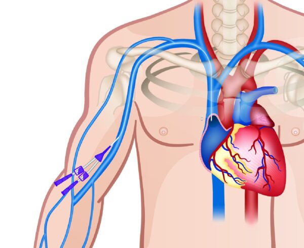
My relative has had angioplasty and has coronary stents. Is it really safe to do an MRI for him? Are joint replacement metallic implants also safe?
Recently done angioplasties make use of MRI safe implants. In the last few years the metallic prosthetic joints are also made of titanium which are MRI safe. Metallic coils used to treat aneurysm are also recently made of MRI safe material and hence do not pose any risk. These may cause artifacts if they are in the area of evaluation and may lead to suboptimal study. However these decisions are taken by the intensivist/ clinician who has ordered the MRI study and the radiologist. It is the responsibility of every patient/ relative to give detailed information of any metallic prosthesis / implant procedure done for the patient.
Is cardiac pacemaker safe within an MRI machine?
The MRI radiations interfere with function of the cardiac pacemaker and hence this information needs to be given to the doctor/ radiologist. The engineer from the respective company of pacemaker is called, he disables the pacemaker temporarily and restarts it after the scan.


Ask a Question?